
Lu Fan, Ole Jung, Markus Herrmann, Marina Shirokikh, Sanja Stojanovic, Stevo Najman, Fabian Körte, Xin Xiong, Katja Schenke-Layland,* and Mike Barbeck
Collagen crosslinking employing ultraviolet A rays and riboflavin (UVA/R) has emerged as a pivotal technique in clinical therapies, especially in ophthalmology since the 1990s. Despite its clinical adoption, the lack of clarity of the detailed mechanism and the imperative for a refined manufacturing process necessitates further investigation. This study advances the understanding of UVA/R crosslinked collagen, concentrating on identifying the primary crosslinking sites using seven synthetic peptides and exploring the pathways of riboflavin.mediated crosslinking.
The results demonstrate that tyrosine residues are key crosslinking sites, and riboflavin plays a dual role as both a catalyst and a competitive inhibitor in the crosslinking process. Furthermore, the UVA/R crosslinked collagen matrix exhibits a more harmonious balance between stability and degradability compared with chemically crosslinked collagen matrices, coupled with superior mechanical properties and augmented biocompatibility. In vivo experiments further.
1. Introduction
Collagens have been extensively utilized in various biomedi- cal fields as functional biomaterials, including wound healing,bone grafts, and regeneration, ophthal- mology, cardiovascular, and neural tissue engineering. To optimize the func- tionality of the collagens, mainly collagen type I (collagen), in these applications, crosslinking is often imperative, enhancing its mechanical properties, biostability, bio- compatibility, and other functionalities. Among the various crosslinking tech- niques, ultraviolet A rays and riboflavin (UVA/R) crosslinking stands out as a particularly promising technique, hav- ing been approved for clinical use in ophthalmological therapy since the early 1990s. This inherent convenience and safety hold tremendous potential for expanding collagens’ application land- scape across diverse biomedical fields.
The realm of UVA photocrosslinking is rapidly advancing, revealing promising ap- plications across medical disciplines. Its utility now extends to creating innovative hydrogels aimed at minimizing postopera- tive adhesions, which has potential to rev- olutionize laparoscopic surgery recovery. Moreover, UVA/R crosslinked acellular porcine cornea are be- ing evaluated as a potential scaffold for Boston Keratopros- thesis, reflecting significant strides in biocompatibility and efficiency. This technique’s application has diversified, with groundbreaking methods like the glutaraldehyde-free prepara- tion for cardiac implants, meniscus tissue engineering,controlled drug delivery device, and extends its influence into cutting-edge research domains including 3D bioprinting and organ-on-a-chip systems, showcasing the dynamic potential of UVA/R crosslinking in modern biomedical engineering.
Despite these promising applications, the mechanism un- derlying UVA/R crosslinking remains an area of ongoing re- search and debate. Many studies suggest that riboflavin functions primarily as a photocatalyst generating reactive oxygen species (ROS) to induce collagen oxidation and polymerization.Alternative theories propose that this is a much more com- plex photochemical process, which is oxygen and/or UVA exposure-dependent. Debates also exist regarding the spe- cific crosslinking sites on collagen molecules, with some assert- ing non-selectivity, while others point to amino acids like ly- sine, histidine, arginine, tyrosine, or methionine.
These dif- fering viewpoints on UVA/R crosslinking mechanisms under- score a lack of comprehensive understanding, presenting chal- lenges in standardizing manufacturing processes and in the reg- ulatory evaluation of medical devices. To address these knowl- edge gaps and potentially increase applications using collagen, our study investigates the fundamental principles of UVA/R crosslinking.
By exploring alternative mechanistic explanations and unraveling the intricate interactions between collagen and ri- boflavin, we provide valuable insights that will contribute to the refinement and optimization of UVA/R crosslinking techniques. However, investigating the crosslinking mechanism in collagen presents significant challenges due to the dynamic photochem- ical processes and collagen molecular complexity.To un- ravel these detailed mechanisms, advanced experimental tech- niques and comprehensive analytical methods are required to identify and characterize the transient intermediates and path- ways accurately. Additionally, the variability in collagen’s physical and chemical properties, influenced by the collagen source, type, processing, and environmental factors add layers of difficulty to these studies, necessitating a nuanced approach to fully understand the crosslinking process.
In our study, we adopted an innovative experimental frame- work, synthesizing seven synthetic peptides representing cru- cial potential crosslinking sites on collagen, focusing on amino acid residues such as tyrosine (Y), lysine (K), arginine (R), me- thionine (M), histidine (H), tryptophane (W), and phenylalanine (F). These amino acids were selected for their reactive groups and positions in the non-helical region of the collagen molecule, which is more accessible for crosslinking reactions and crucial for enhancing mechanical properties. (Figure 1) By exposing these peptides and native collagen to varied UVA energy and ri- boflavin ratios, we dissected the crosslinking process using liquid chromatography-mass spectrometry (LC-MS). We also compared UVA/R crosslinking with chemical crosslinkers, utilizing Raman microspectroscopy to discern distinct molecular alterations post- crosslinking. We evaluated mechanical strength, thermal stabil- ity, degradation resistance, and cytotoxicity, culminating in in vivo implantation trials to assess biocompatibility and tissue re- generation potential.
By dissecting the crosslinking process at a molecular level, our study reveals critical insights into the roles and interactions of specific amino acids, shedding light on the in- tricate dynamics of UVA/R crosslinking. These discoveries hold substantial implications for advancing biomaterial science, oering a foundation for developing advanced, biocompatible, and mechanically robust biomaterials, thus paving the way for more effective and safer biomedical applications.
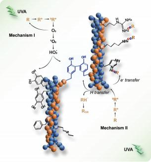
Figure 1. Schematic of UVA/riboflavin collagen crosslinking. UVA: Ultra- violet A light, the source of photoactivation. R: ground state riboflavin. R*: excited state of riboflavin upon initial UVA exposure. 3 R*: Triplet state ri- boflavin after intersystem crossing, a higher energy state capable of engag- ing in crosslinking reactions. O2: molecular oxygen. 1 O2: singlet oxygen, a reactive oxygen species generated by energy transfer from photoactivated riboflavin to molecular oxygen. HO2 •: hydroperoxyl radical. RH: general structure representing a hydrogen donor. Rox : oxidized form of the hy- drogen donor following hydrogen transfer. Amino acids: Y (tyrosine), K (lysine), R (arginine), M (methionine), H (histidine), W (tryptophan, not present in porcine collagen type I), F (phenylalanine).
2. Results and Discussion
2.1. Preparation and Characterizations ofthe Collagen Matrix
Type I collagen, sourced from porcine skin due to its close re- semblance to human tissue, oers a robust framework for tissue engineering.This form of collagen is ideal for biomedical applications because of its abundance in the skin and its well- defined triple-helical structure comprising two α1 chains and one α2 chain (Figure2A). SDS-PAGE indicated the integrity and purity of our porcine-derived collagen after a series of me- chanical homogenization processions, using rat tail collagen as a control.
Figure 2B showcases distinct bands in the SDS-PAGE gel for both porcine skin-derived collagen (P-col) and rat tail- derived collagen (R-col), highlighting the α 1 and α2 bands at ≈150 and 140 kDa, respectively. No further bands below the 100 kDa were observed. Higher molecular weight bands, corresponding to dimers (β) and trimers (y), were also discernible, indicating the collagen matrix’s high purity and maintained integrity following mechanical processing.Circular dichroism (CD) spectroscopy provided further confirmation, exhibiting characteristic negative and positive peaks that signify the presence of the collagen’s well- maintained triple-helical conformation (Figure2C).Such high purity and structural integrity are crucial for the effective interac- tion with crosslinking agents and precise mechanical property analysis.
Scanning electron microscopy (SEM) was employed to eval- uate morphological changes in the collagen matrices before and after UVA/R crosslinking. (Figure 2D) Initially, the non- crosslinked collagen displayed a unique crystal-like surface tex- ture in its hydrated-frozen state, deviating from the conven- tional fibrillar structure due to the homogenization and stir- ring/foaming processes implemented during its preparation (Figure 2Da). This intentional disruption was necessary for fur- ther matrix design. Following crosslinking, a noteworthy trans- formation was observed in the UVA/R crosslinked collagen matrix. It exhibited an intricate, porous architecture with inter- connected voids and pores, ranging from 1 to 20 μm in size, contributing to its three-dimensional complexity. (Figure 2Db) The heterogeneity in pore size may foster a more natural and dynamic environment for cells, allowing for varied cell adhesion, migration, and nutrient diusion, which are crucial for tissue re- generation and repair.Moreover, the restructured collagen fibers may enhance mechanical cues that can guide cellular align- ment and dierentiation, thus contributing to the formation of a more functional and organized tissue structure in biomedical applications.
Furthermore, the densification and increased stiness resulting from lyophilization suggest enhanced resis- tance to mechanical deformation, an essential characteristic for biomaterials exposed to physiological stresses (Figure 2Dc,d). This enhanced mechanical robustness not only ensures the struc- tural integrity of the collagen matrix under varying physical con- ditions but also potentially extends its functional lifespan, mak- ing it more suitable for long-term biomedical applications such as implantable devices or tissue sca olds.
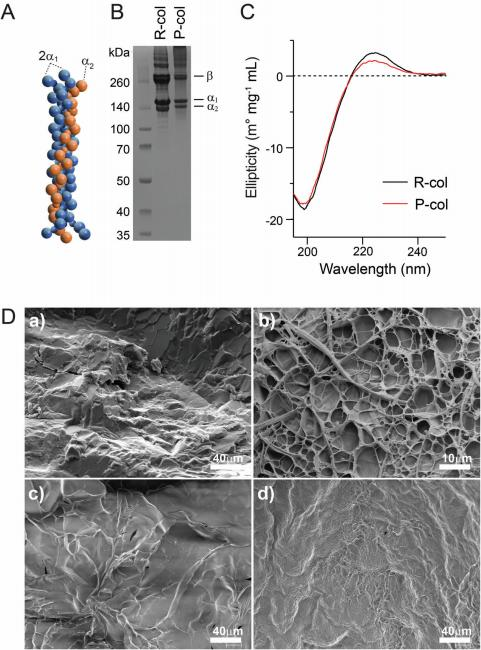
Figure 2. Structural and mechanical characterization of collagen pre- and post-UVA/riboflavin treatment. A) Molecular model of the collage type I triple helix structure, the fundamental building block of collagen matrices. B) SDS-PAGE analysis depicting the characterization of collagen type I extracted from rat tail (R-col) and porcine skin (P-col). C) Circular dichroism (CD) spectra of R-col and P-col, with ellipticity indicating preservation of the colla- gen secondary structure. D) Scanning electron microscopy images presenting the morphological examination of non-crosslinked collagen and UVA/R crosslinked collagen in their hydrated-frozen state (a,b), as well as in their lyophilized state (c,d).
2.2. Exploration of the UVA/R Crosslinking Mechanism
Having established the fundamental aspects of the collagen matrix preparation and analyzed its initial characteristics, we then delved into the exploration of the mechanism of UVA/R crosslinking, a pivotal process that significantly alters and en- hances the matrix’s properties. This section investigates where crosslinks form on the collagen molecule, the role of riboflavin as a catalyst or participator, and further postulates the necessity for adjustments ofthe collagen UVA/R crosslinking in the vari-ous applications.
2.2.1. Where do Crosslinks Form on the Collagen Molecule?
In our study, we probed the crosslinking mechanism of type I collagen at the molecular level, focusing on non-helical regions that enhance mechanical properties post-crosslinking. Employ- ing seven synthetic peptides representing crucial amino acids— tyrosine (Y), lysine (K), arginine (R), methionine (M), histidine (H), tryptophane (W), and phenylalanine (F)—we aimed to iden- tify specific sites of crosslinking. These amino acids were se- lected for their distinct reactive functionalities and strategic po- sitions within collagen the molecule (Figure S1, Supporting In- formation). Specifically, tyrosine (Y) possesses a reactive phenol group capable of engaging in covalent bonding.Lysine (K) and arginine (R) are equipped with amino groups that are can- didates for chemical bond formation.
Histidine (H) is char- acterized by an imidazole ring, and phenylalanine (F) by an aro- matic ring, both oering potential sites for reactivity.Our LC- MS analyses delineated a distinct pattern: modifications post- UVA/R treatment occurred exclusively in tyrosine-bearing pep- tides, likely due to the generation of covalent di-tyrosine link- ages (Figure 3A,B). This specificity was further accentuated by the absence of covalent bonding products in the other six amino acids including peptides, underscoring the preferential role of ty- rosine in the crosslinking mechanism (Figures S2 and S3, Sup- porting Information). The propensity for di-tyrosine formation,initially posited from our synthetic peptide studies, was con- clusively corroborated by the LC-MS analysis of hydrolysates of UVA/R crosslinked collagen (Figure 3C,D).
During the exami- nation of UVA/R crosslinked collagen, di-tyrosine was reliably identified by using multiple reaction monitoring (MRM) mode in mass spectrometry and authenticated through comparisons with a di-tyrosine standard.Notably, our LC-MS scrutiny of collagen hydrolysates disclosed a peak highly akin to, yet dis- tinguishable from, the di-tyrosine standard. Acknowledging the likelihood of isomeric or spatial variations within the collagen molecules, we cautiously infer, predicated on mass considera- tions, that the coinciding peaks could represent di-tyrosine forms (Figure S4, Supporting Information).Given the protein se- quence of porcine type I collagen (Access numbers A0A1S7J210 for COL1A1 and A0A1S7J1Y9 for COL1A2), the complete ma- ture collagen molecule should exhibit a molecular weight of 282.85 kDa, encompassing 3152 amino acid residues, inclusive of 13 tyrosine residues.
If we assume all these tyrosine residues are equally reactive and available for crosslinking, our LC-MS re- sults intimate that a conservative estimate of crosslinked tyrosine within the collagen matrix is at least 14.3% (Figure S5, Support- ing Information). This deduction is instrumental in appraising the extent of collagen crosslinking aected by UVA/R conditions and enriches our comprehension of its mechanisms.
The substantiation of di-tyrosine in hydrolyzed UVA/R crosslinked collagen is a pivotal affirmation of tyrosine’s exclu- sivity in the crosslinking schema. Such evidence not only echoes the synthetic peptide experimental results but also exemplifies the practical relevance of tyrosine crosslinking in collagen matri- ces. The presence of di-tyrosine serves as a molecular hallmark of successful crosslinking, offering an imperative insight that is anticipated to be crucial for refining UVA/R crosslinking tech- niques in collagen tailored for biomedical utilizations.
2.2.2. Riboflavin: Crosslinking Catalyzer or Participator?
In exploring the role of riboflavin in collagen crosslinking, our study uncovers its dual function as both a catalyst and a par- ticipator. By examining the relationship between tyrosine and riboflavin ratios in both the peptide and collagen systems, we observed similar notable trends: a positive correlation between crosslinking ecacy and riboflavin at lower ratios, which reverses at higher concentrations (Figure 3E). For example, in collagen, at lower riboflavin concentrations (tyrosine/riboflavin ratios of 1/0.0625 to 1/0.25), there was a significant increase in the stor- age modulus compared to the non-crosslinked collagen. How- ever, at higher concentrations, the storage modulus sharply de- clined (Figure 3F).
The noticeable reduction in the mechanical strength of col- lagen matrices at elevated riboflavin concentrations prompted an exploration of the underlying mechanisms. Our investiga- tion was guided by a theoretical kinetic model, indicating a criti- cal dependence of the two pathways of riboflavin-mediated pho- tocrosslinking on ambient oxygen levels.This model inspired our discovery: the crosslinking efficiency is markedly sensitive to riboflavin concentration, particularly in environments with con- sistent oxygen availability.
This nuanced photochemical behav- ior of riboflavin suggests two primary reaction pathways upon UVA excitation, as illustrated in Figure 1. Initially, riboflavin acts as a photocatalyst, generating ROS that facilitate intermolecular crosslinking, predominantly at tyrosine residues located in the non-helical regions of collagen. This process enhances the me- chanical properties of the matrix. However, an alternative path- way becomes predominant at higher riboflavin concentrations, where riboflavin actively engages in the crosslinking reaction, depleting ROS and subsequently diminishing crosslinking effi- ciency. Furthermore, electron transfer processes may induce in- teractions between positively charged amino acid residues like lysine, histidine, and arginine, and negatively charged riboflavin, potentially influencing the mechanical properties of the collagen matrix. This aspect merits further experimental validation.
Additionally, UVA exposure for crosslinking the collagen ma- trix was also optimized in our study. Our findings as illustrated in Figure 3E indicate that the optimal UVA exposure for colla- gen matrix crosslinking was 10800 mJ cm_2. Beyond this UVA dosage, the storage modulus of the collagen matrices noticeably decreased, suggesting potential degradation of the collagen scaf- fold due to overexposure. Interestingly, for peptide crosslinking, the optimal UVA dosage was markedly lower, at 600 mJ cm_2 (Figure S6, Supporting Information); and in another work, Ue- mura et al. identified that the optimal UVA exposure for human dentin collagen crosslinking was 1600 mJ cm_2.
Overall, these insights uncover an additional layer of the UVA/R crosslinking mechanism, highlighting the crucial need to fine-tune riboflavin concentration and UVA exposure for prac- tical applications. The notable disparity between in vivo tissue environments and in vitro collagen crosslinking systems is sub- stantial. Variations in collagen sources (tyrosine residue content), concentration, density, and differing buffer conditions necessi- tate tailored approaches for adjusting riboflavin concentration and UVA exposure to achieve optimal crosslinking outcomes.
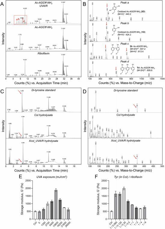
Figure 3. Dissecting the UVA/R crosslinking mechanism in collagen. A) LC-MS chromatograms post UVA/R treatment of Ac-AGGY-NH2 peptide with control samples of Ac-AGGY-NH2 and riboflavin. B) Mass spectra correlating with newly emerged peaks (a–d) in chromatograms of UVA/R treated Ac-AGGY-NH2. C) LC-MS chromatograms of di-tyrosine standard and hydrolysates of non-crosslinked collagen and UVA/R crosslinked collagen, D) with corresponding mass spectra. E) Storage modulus (G’) of collagen as a function of UVA exposure (mJ cm_2 ), showcasing the dose-dependent increase in mechanical properties. F) Storage modulus (G’) in relation to the Tyr (in collagen)/riboflavin ratio, illustrating the impact of varying riboflavin concentrations on the mechanical strength of the collagen matrices.
2.3. Comparative Investigation of the Impact of UVA/R and Chemical Crosslinking on Collagen Matrices
After elucidation of the complexity of the UVA/R crosslinking mechanism, we turned our attention to the comparative investi- gation aimed at comprehensively evaluating the effects of differ- ent chemical crosslinking techniques on collagen matrices. Each analysis contributes to a holistic understanding of the structural and functional alterations induced by UVA/R and other chemical crosslinking methods.
2.3. 1. Raman microspectroscopy
Raman microspectroscopy has offered a profound insight into the molecular integrity of collagen matrices subjected to different crosslinking techniques.Detailed peak assignments along with their corresponding references are compiled in Table S3 (Supporting Information). In our study, Col UVA/R crosslinked collagen (Xcol_UVA/R) and EDC/NHS crosslinked collagen (Xcol_EDC/NHS) retained their quintessential secondary struc- tures post-crosslinking when compared with non-crosslinked collagen (Col), as evidenced by the consistent amide bands across all samples: Amide III (1260 cm_1), Amide II (1452 cm_1), and Amide I (1664 cm_1)(Figure4A).
The spectra revealed that while the characteristic triple-helix structure of collagen was main- tained, there were notable reductions in the intensity of peaks (815, 936, 971, and 1080 cm_1) associated with the side-chain vi- brations of amino acids involved in both UVA/R and EDC/NHS crosslinking reactions. Such findings suggest subtle yet definitive molecular rearrangements within the collagen’s ar- chitecture, particularly aecting vibrational modes, such as C- C stretching, C-C stretching wagging, and CH3 rocking within lysine, hydroxylysine, and tyrosine residues, which are crucial for the crosslinking process.
Notably, the extent of intensity re- duction was more pronounced in EDC/NHS crosslinked colla- gen, indicating a higher abundance oflysine and hydroxylysine- mediated crosslinks compared to the tyrosine-mediated ones in UVA/R crosslinking. An intriguing shift from 936 to 927 cm_1 after EDC/NHS crosslinking possibly alludes to heightened ten- sion arising from proline residue packing post-crosslinking.
Further scrutiny via multivariate principal component analy- sis (PCA) corroborated these structural nuances, discriminating between the untreated and crosslinked samples (Figure 4B–D). The distinct clustering of PC-3 and PC-4 scores elucidated the molecular underpinnings unique to each treatment, with the UVA/R crosslinking manifesting a distinct molecular fingerprint when compared to its chemical counterpart. The PC-3 scores of- fered positive scores evident for Xcol_UVA/R spectra and neg- ative scores for Col and Xcol_EDC/NHS. This trend was mir- rored in the PC-3 loading plot, featuring prominent peaks at 936 cm_1 (proline) and 1688 cm_1 (Amide I).
In tandem, the PC-4 scores revealed positive ranges for both Xcol_UVA/R and Xcol_EDC/NHS, whereas Col spectra clustered in the nega- tive range. The corresponding PC-4 loadings plot pinpointed crosslinking-associated peaks at 896, 980, 1099, 1309, 1439, and 1655 cm_1, signifying the vibrational modes of0 (C-C), 0 (C-N), as well as CH3 /CH2 twisting, bending, or wagging mode deforma- tion among others, reinforcing the crosslinking-driven molecular distinctions observed.
Raman microspectroscopy provided molecular fingerprinting patterns that elucidate the changes occurring at the molecular level in collagen post-crosslinking, as well as the different charac- teristics between collagen crosslinked by UVA/R and EDC/NHS methods. These spectroscopic findings are pivotal in guiding the functionalization of collagen for advanced biomedical applica- tions.
2.3.2. Size-Exclusion Chromatography of Digested Collagen Matrix
Size-exclusion chromatography (SEC) of collagenase-digested samples revealed marked differences in the molecular profile of collagen matrices post-crosslinking (Figure 4E–G). Given that collagenase primarily cleaves the peptide bonds within collagen molecules at the characteristic Gly-X-Y triplets, where X and Y are often proline or hydroxyproline residues, the enzymatic di- gestion process yields smaller peptide fragments. The presence of larger peptide fragments in crosslinked samples, especially noticeable in the region of peak 1, is indicative of the effective- ness of crosslinking in impeding enzymatic cleavage. This find- ing was particularly pronounced in the UVA/R crosslinked sam- ple, where the sizeable peptide entities were retained to a degree.
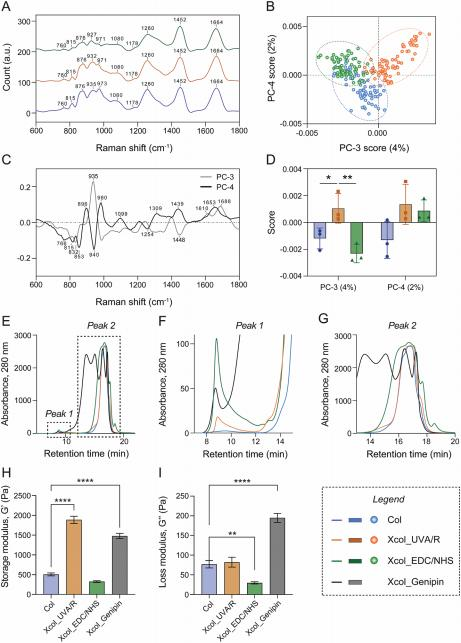
Figure 4. Comprehensive characterization of non-crosslinked collagen (Col), UVA/R crosslinked collagen (Xcol_UVA/R), EDC/NHS crosslinked collagen (Xcol_EDC/NHS), and genipin crosslinked collagen (Xcol_Genipin) matrices. A–D) Raman microspectroscopic analysis: (A) Average Raman spectra highlighting distinct peaks and patterns. Each condition was replicated at least thrice (n ≥ 3), with multiple spectra acquired for each replicate (n ≥ 25). Multivariate data analysis outcomes include principal component analysis (PCA) results featuring PCA spectral loadings of PC-3 and PC-4 (C), scores plot of PC-4 versus PC-3 (B), and statistical representation of PC-3 and PC-4 scores as mean values ± SD (D). Confidence ellipses in (B) signify 95% confidence intervals. E–G) Size-exclusion chromatography analysis: traces depicting the behavior of collagen matrices, as detected using a UV detector. Close-up views of Peak 1 and Peak 2 are detailed in (F) and (G). Dynamic H) storage modulus (G’) and I) loss modulus (G’’) extracted from the plateau LVE region during frequency sweeps.
similar to that of chemically crosslinked samples. Such an ob- servation highlights the comparable efficacy of UVA/R crosslink- ing to traditional chemical methods like EDC/NHS and genipin in protecting collagen matrices from enzymatic digestion. Moreover, the UVA/R crosslinking demonstrated an advanta- geous profile with fewer smaller by-products relative to chem-ical crosslinking.
The chromatograms for the EDC/NHS and genipin-treated samples showed additional peaks, suggesting the presence of by-products, which could potentially lead to cytotoxic eects or provoke immune responses when used in biomedical applications.In contrast, the UVA/R crosslinking process appears to have resulted in a cleaner profile with less fragmentation, which may translate to a more favorable safety profile for in vivo applications. The molecular integrity main- tained by UVA/R crosslinking, coupled with its reduced by- product profile, positions it as a potent method for preparing collagen-based biomaterials for regenerative medicine.
2.3.3. Rheological Properties
The rheological properties of crosslinked collagen matrices, as analyzed by dynamic shear-stress amplitude and frequency sweeps, provided insights into the mechanical implications of each crosslinking strategy at physiological temperatures. (Figure 4H,I) Xcol_UVA/R demonstrated an enhanced elastic profile as evidenced by its storage modulus, without a corre- sponding loss in material resilience. This balance of elasticity and durability underscores the potential of UVA/R crosslinking to fortify the mechanical integrity of collagen scaffolds without sacrificing the material’s ability to absorb and dissipate energy, which is an essential characteristic for biomaterials that must en- dure dynamic physiological environments.
On the other hand, Xcol_EDC/NHS presented an unexpectedly lower storage modu- lus (Figure 4I), suggesting that while chemical crosslinking with EDC/NHS does modify the material, it may not uniformly en- hance the elastic properties of the collagen matrix. This find- ing could have implications for the application of EDC/NHS crosslinked materials in scenarios where mechanical stiness is a prerequisite. In contrast, Xcol_Genipin showed a marked in- crease in stiness, as indicated by its higher storage modulus. However, the increased rigidity may impose limitations on the material’s functional compatibility, particularly concerning cel- lular activities. Materials that are excessively sti could poten- tially hinder cell migration and proliferation, both of which are critical for tissue regeneration and integration.
Taken together, these results suggest that UVA/R crosslinking achieves an optimal balance of stiffness and flexibility, which may be conducive to cellular functions, thus presenting a promising avenue for further development of collagen-based scaolds for regenerative medicine.
2.3.4. Thermal Stability
Dierential scanning calorimetry (DSC) measurements, de- picted in Figure 5A,B, provided essential insights into the ther- mal stability of the collagen matrices. The denaturation tempera- ture ( Tm), observed as a peak on the DSC curves, marks the tran- sition point where the collagen triple helix begins to unwind due to the breakdown of stabilizing hydrogen bonds.This thermal event is accompanied by the absorption of heat, which is quantita- tively measured as the change in enthalpy (ΔH).
In our study, a noticeable elevation in Tm was recorded for the crosslinked collagen samples. Specifically, Xcol_UVA/R and Xcol_EDC/NHS show Tm values at 75 and 76.0 °C respectively, indicating en- hanced thermal stability potentially conducive to maintaining structural integrity at physiological temperatures. Xcol_Genipin, while displaying a lower Tm at 60.1 °C, still shows increased thermal resistance relative to non-crosslinked collagen. The en- thalpy changes (ΔH) associated with the denaturation process,as depicted in Figure 5B, further support these findings, with Xcol_UVA/R and Xcol_EDC/NHS demonstrating a substantial absorption of heat, reflective of the energy required to disrupt the strengthened collagen structure due to crosslinking. These thermodynamic characteristics imply that the crosslinking con- fers additional stability to the collagen matrices, which is crucial for their performance under thermal stress in vivo.
2.3.5. Degradation Resistance
Our study extended to a comparative degradation analysis of crosslinked collagen matrices in physiologically relevant condi- tions using phosphate-buffered saline (PBS) and collagenase so- lutions (Figure 5C,D). The degradation profile of Xcol_UVA/R was particularly compelling, exhibiting resistance on par with chemically crosslinked matrices, thereby marking a significant improvement over the non-crosslinked collagen. This observa- tion is pivotal, as it is the first to directly compare the degra- dation resistance of UVA/R crosslinked collagen with chemi- cally crosslinked variants. Xcol_EDC/NHS displayed exceptional durability in PBS, maintaining over 60% of its mass after 30 days, indicative of its potential for applications where extended stability is paramount. Genipin-crosslinked collagen also showed remark- able resistance in collagenase solutions, suggesting its suitability for environments with active enzymatic degradation.
However, the robustness of these chemically crosslinked ma- trices raises concerns in the tissue engineering context, where scaffold degradation kinetics must be synchronized with tissue regeneration.[75] Biomaterials that degrade too slowly may dis- rupt the delicate process of new tissue formation, potentially in- terfering with cell migration, vascularization, and the natural tis- sue remodeling process.[30] Moreover, the persistent presence of non-degraded materials can provoke a sustained inflammatory response, which may adversely affect healing outcomes.[31] In tis- sue engineering, the ideal biomaterial should provide initial me- chanical support but gradually cede its place to newly formed tis- sue. Therefore, optimizing the degradation rate to ensure a seam- less transition from scaffold to native tissue is critical. Our find- ings highlight the need for a nuanced approach to the design of collagen-based scaffolds, aiming for a delicate equilibrium that supports initial tissue repair and subsequently allows for natu- ral tissue integration without eliciting chronic inflammation or other negative responses.
2.3.6. In Vitro Cytotoxicity Test
The biocompatibility of our collagen matrices was evaluated through the CCK-8 assay utilizing L929 fibroblasts, following DIN EN ISO 10993–5: 2009 and DIN EN 10993-12: 2021 guide- lines. The standard posits that biomaterials should not reduce cell viability below 70% of the control to be considered biocompatible. As shown in Figure 5E, the crosslinked collagen matrices, partic- ularly Xcol_EDC/NHS, exhibited reduced cell viability, suggest- ing potential cytotoxicity from residual crosslinking agents or by- products. Contrastingly, Xcol_UVA/R surpassed the biocompati- bility threshold with viability above 80%, indicating its suitability for tissue engineering applications due to its favorable balance of stability and degradation.
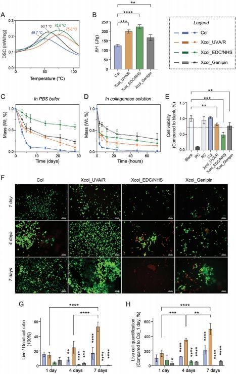
Figure 5. Characterization of collagen matrices and in vitro biological evaluation. A) Differential scanning calorimetry (DSC) curves illustrating the thermal transitions of collagen matrices, marked with peak temperatures (Tm). B) Enthalpy changes (ΔH) during denaturation of collagen matrices. Mass loss during in vitro degradation in C) phosphate-buffered saline (PBS) solution and D) collagenase solution. E) In vitro cytotoxicity test using L929 fibroblasts via CCK-8 assay, following the ISO 10993–5 standard. F) Representative confocal laser scanning microscopy (CLSM) images of live- dead (Calcein-PI) staining of L929 cells cultured on collagen scaffolds at 1-, 4-, and 7-days post-seeding. G) Quantitative analysis of live/dead cell ratio, derived from the CLSM images, using ImageJ software for cell quantification. H) Quantification of live cells, highlighting the cell viability and proliferation across the study period. Statistical evaluation for parts (G) and (H) was performed using ANOVA for multiple comparisons at different time points, with specific comparisons made between the Xcol_UVA/R group and other groups. (Scale bars in the live-dead staining images correspond to 100 μm. Calcein in green, PI in red.).
Live-dead staining (Figure 5F) with its subsequent quantita- tive analysis (Figure 5G,H) further highlighted the superior per- formance of Xcol_UVA/R, which supported cell survival and proliferation. Initially, at day 1, the live/dead ratios for Col and Xcol_UVA/R groups were comparable, corroborating the results of the extraction cytotoxicity test depicted in Figure 5E. However, as the culture progressed to days 4 and 7, the Xcol_UVA/R scaf- fold demonstrated its superiority with significantly enhanced cell viability and proliferation, likely attributable to its biomechanical attributes. While the non-crosslinked collagen scaffold, despite being biocompatible, did not exhibit the structural integrity nec- essary for optimal cell-material interactions, as evidenced by its performance at 4 and 7 days. In contrast, the collagen matrices crosslinked with EDC/NHS and genipin displayed diminished live cell ratios throughout the duration of the study, which could raise concerns regarding their cytotoxicity profiles.
Our comprehensive analyses encompassing enzymatic diges- tion profiles, mechanical strength, thermal stability, and degrada- tion resistance, when juxtaposed with in vitro cytotoxicity assess- ments, reveal a multifaceted evaluation of crosslinked collagen matrices for biomedical applications. Xcol_UVA/R stands out with its balanced profile; it exhibits comparable thermal stabil- ity and degradation resistance to chemically crosslinked counter- parts, maintaining structural integrity under physiological con- ditions while supporting robust cell viability and favorable cell- material interactions.
The enzymatic digestion results, indicat- ing reduced degradation in crosslinked samples, align with the observed enhancement in mechanical and thermal properties, corroborating the crosslinking’s effectiveness in bolstering ma- trix resilience. However, the mechanical robustness and reduced degradability, as seen inXcol_EDC/NHS and Xcol_Genipin, may be a detriment to the delicate equilibrium required for tissue re- generation. These matrices show limited enzymatic breakdown and increased thermal resistance, yet this durability could po- tentially impede natural tissue remodeling, as indicated by their lower cell viability.
In the realm of tissue engineering, where scaf- fold degradation is expected to pave the way for new tissue for- mation, materials like Xcol_UVA/R that strike an optimal bal- ance between stability and degradability are preferred.They facilitate cellular functions without invoking a prolonged inflam- matory response, thereby enhancing the healing outcomes.In summary, the choice of crosslinking method profoundly impacts the scaffold’s performance, with UVA/R crosslinking emerging as a promising approach to creating biocompatible, stable, yet degradable scaffolds conducive to successful tissue engineering applications.
2.4. Histological Assessment of Subcutaneous Implants
The exceptional biocompatibility and osteoconductivity of colla- gen make it a cornerstone for bone grafting applications. How- ever, its utility has historically been limited by biomechanical sta- bility. Through the application of an innovative UVA/R crosslink- ing process, we have successfully addressed this limitation, sig- nificantly enhancing the mechanical stability of our collagen scaf- folds. The integration of inorganic bone substitutes (BS), such as hydroxyapatite and β-TCP, provides the necessary mechan- ical support for long-term bone remodeling, while lacking the ECM components crucial for early cell interactions and scaf- fold degradability.
Our innovative injectable composite scaffold with UVA/R crosslinked collagen matrix and BS, therefore, is designed to optimize both initial cell engagement and endur- ing bone remodeling. Through subcutaneous evaluationsand de- tailed histological assessments ofthe implanted scaffolds, we’ve achieved a nuanced understanding of material integrity and tis- sue response dynamics. This includes an in-depth analysis of tissue reactions, notably immune responses, to ascertain bio- compatibility and derive critical insights for future applications. This methodical approach underscores our commitment to thor- oughly exploring the interaction between our engineered matri- ces and biological systems.
2.4. 1. Tissue Reaction Analysis
Movat’s Pentachrome staining is a multipurpose histological technique that distinctly marks various components of con- nective tissue, facilitating their differentiation in histological sections.In this study, Movat’s pentachrome staining was uti- lized to visually distinguish and semi-quantitatively analyze GAG and collagen distribution within the tissue sections, revealing the distinct behaviors ofthe implanted non-crosslinked collagen- and UVA/R crosslinked collagen-based scaffolds (BS-Col and BS- Xcol_UVA/R) within the tissue environment (Figure 6A–C). By day 10 post-implantation, both groups showed collagen matri- ces within the implantation beds (Figure 6B).
Notably, the BS- Xcol_UVA/R group demonstrated a remarkable adherence to the bone substitute surface in contrast to the loosely associated ma- trix observed in the BS-Col group, suggesting enhanced matrix stability potentially conferred by the UVA/R crosslinking process. The quantitative analysis further corroborated these findings, in- dicating a higher collagen content within the BS-Xcol_UVA/R implantation sites compared to BS-Col (Figure 6C), which may proof the profound effect of crosslinking on matrix integrity. As the tissue formation progressed, the initially similar gly- cosaminoglycan (GAG) levels took a consequential turn, show- ing a decrease over time, suggesting an ongoing tissue response, cellular activity, and a dynamic matrix remodeling process over time.
By day 30, the BS-Xcol_UVA/R group exhibited a striking re- tention of collagen content, with the matrix persisting around the bone substitute surfaces. This sustained presence, validated by quantitative analysis, highlighted the enduring effects of UVA/R crosslinking on matrix stability. (Figure6C) The tissue within the scaffolds had matured, as evidenced by the increased vascular- ization in both groups. However, a distinct immune profile was observed; the BS-Col group showed a higher prevalence of macrophages and multinucleated giant cells, suggesting an on- going response to the scaffold degradation.
2.4.2. Immune Response Evaluation
Our histological exploration into the immune responses at the subcutaneous implantation sites revealed nuanced interactions between the host’s defenses and the implanted collagen matrix- based composites. Initially, at day 10, pro-inflammatory CD11c- positive macrophages were prominent around the implants.
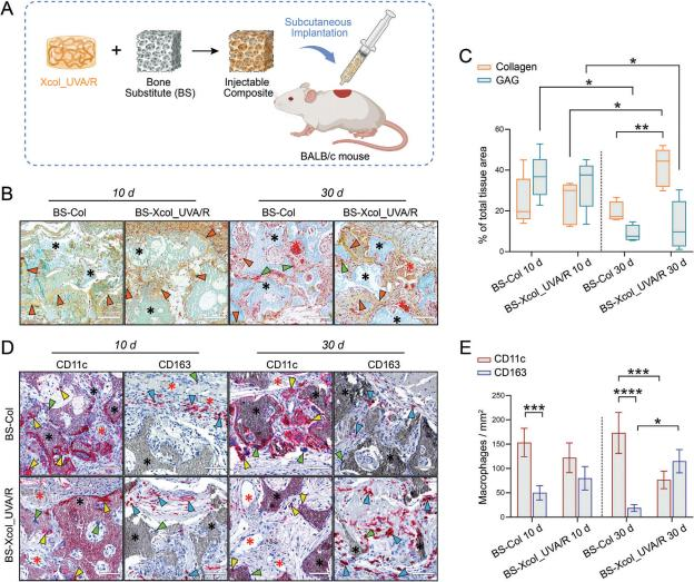
Figure 6. In vivo tissue reaction and immune response to collagen matrix-based materials: BS-Col and BS-Xcol_UVA/R. A) Schematic illustration depicting the injectable composite material composed of collagen matrix and bone substitute (BS), subcutaneously implanted into BALB/c mouse.
B) Representative histological images ofthe implantation areas of BS-Col and BS-Xcol_UVA/R at day 10 and 30 post-implantation using Movat’s Pen- tachrome staining. C) Quantification of Movat’s Pentachrome via color deconvolution analysis using ImageJ. Yellow-green = collagen, green-blue = glycosaminoglycan (GAG). Presented as a percentage of the total stained area. D) Representative immunohistochemical images of pro-inflammatory (CD11c) and anti-inflammatory (CD163) macrophage subtypes in the implantation areas of the respective groups. E) Histomorphometric (quantitative) analysis of CD11c-positive and CD163-positive macrophage subtypes in the implantation areas of the respective groups using ImageJ. Black stars = bone substitute (BS), red stars = newly formed vessels, orange triangle = collagen, green triangle = multinuclear giant cells, yellow triangle = CD11c-positivie cells, blue triangle = CD163-positive cells. Scale bar = 100 μm.
In both groups, with fewer CD163-positive anti-inflammatory macrophages present in the periphery (Figure 6D). Quantitative assessments (Figure 6E) further delineated the immune land- scape, showing a predominance of M1 macrophages (CD11c- positive) over M2 macrophages (CD163-positive) in the BS-Col group, with cell counts of 153.3 ± 29.0 cells/cm2 and 50.0 ± 14.7 cells/cm2 , respectively. In contrast, the BS-Xcol_UVA/R group exhibited fewer M1 macrophages (122.0 ± 30.5 cells/cm2 ) and a higher incidence of M2 macrophages (79.9 ± 24.2 cells/cm2 ), suggesting a moderated pro-inflammatory response.
By day 30, a discernible shift in macrophage polarization was observed. The BS-Col group maintained to show strong CD11c expression (173.0 ± 42.1 cells/cm2 ) within the implant vicinity,with scant CD163 expression. In contrast, the BS-Xcol_UVA/R group demonstrated a markable increase in anti-inflammatory CD163-positive macrophages (115.0 ± 23.9 cells/cm2 ) and a re- duction in CD11c expression (76.5 ± 17.9 cells/cm2 ), as quanti- fied in Figure 6E. This shift signifies a significant tilt toward an anti-inflammatory phenotype in the BS-Xcol_UVA/R group, with a striking departure from the persistent pro-inflammatory milieu in the BS-Col group.
The histological assessment of subcutaneous implants pro- vides compelling evidence of the UVA/R crosslinking pro- cess’s efficacy in enhancing the biostability and integration of collagen matrices within a biological milieu. Early-stage pro-inflammatory macrophage presence, transitioning to an anti-inflammatory profile in the BS-Xcol_UVA/R group, indi- cates a typical acute immune response, essential for wound heal- ing and implant integration.The reduced degradation prod- ucts and the presence of oxidized tyrosine (DOPA) crosslinked collagen post UVA/R treatment likely contribute synergistically to anti-inflammatory effects. DOPA, which was also one of the products in tyrosine-contained peptides post UVA/R treatment (Figure 3B), plays a significant role in mitigating inflamma- tion, potentially enhancing the biocompatibility of the colla- gen scaffolds.
This opens up avenues for further research into photochemically crosslinked biomaterials in tissue engineer- ing and regenerative medicine, particularly in understanding and harnessing these mechanisms for controlled immune re- sponses in clinical implant integration and functionality. While our study provides comprehensive insights into biocompatibil- ity and mechanical properties, future research incorporating second harmonic generation (SHG) analysis could offer addi- tional understanding of the ultrastructural changes in colla- gen fibrillarity,enriching our exploration of photochemi- cally crosslinked collagen for tissue engineering and regenerative medicine.
The optimized UVA/R crosslinking process enhances our col- lagen scaffolds’ integration with BS materials, creating a highly effective solution for bone grafting. This innovative approach not only strengthens biomechanical stability but also maximizes the biocompatibility of the composite material, making it well- suited for clinical applications. By incorporating inorganic mate- rials into the collagen matrix, we address the essential need for durable mechanical support, pivotal for successful bone regen- eration and remodeling. Crucially, our method boosts initial cell- scaffold interactions, a key element in tissue engineering, while providing the mechanical support vital for bone regeneration.
Beyond bone regeneration, the versatility of UVA/R crosslinked collagen extends to a broad range of applications. It serves as a foundational material for cartilage repair, where its ability to support chondrocyte adhesion and proliferation can be critical. In wound healing, its excellent biocompatibility and con- ducive environment for cell migration accelerate tissue repair.
For vascular tissue engineering, the scaffold supports endothelial and smooth muscle cell growth, essential for forming functional blood vessels. In nerve regeneration, the scaffold can guide neurite outgrowth, offering a promising avenue for repairing nerve injuries. Additionally, the material’s tailored degradation rates and compatibility with various bioactive agents make it an excellent candidate for targeted drug delivery and controlled release systems.
To unlock the full potential of these materials in clinical settings, a deeper understanding of the underlying mechanisms is essential. Advanced imaging and spectroscopy technologies will play a crucial role in this exploration, offering insights into the scaffold’s interaction with biological tissues and its impact on cell behavior. This deepened understanding is critical for tailoring the materials to meet the specific demands of various regenerative medicine applications, ensuring optimized outcomes for tissue repair and regeneration.
3. Conclusion and Outlook
The intricate interplay between the complexity of the collagen molecule and the mysterious UVA/riboflavin (UVA/R) photo-chemical cross-linking process is a fascinating puzzle that our study has skillfully unraveled. Our comprehensive investigation into the crosslinking mechanism of collagen using six strate- gically selected synthetic peptides has yielded remarkable in- sights. HPLC, LC-MS consistently unveil a preference for ty- rosine among the amino acids, a revelation that significantly advances our comprehension of the widely employed UVA/R crosslinking technique, particularly in ophthalmology.
The pref- erential targeting of tyrosine sheds light on the specific molecu- lar pathways involved in UV-induced collagen stabilization. Our findings underscore the role of tyrosine in UV-induced crosslink- ing and the phenol group in tyrosine is also known to be sus- ceptible to photoactivation offering potential engineering strate- gies for artificial or functionalized biomaterials. Beyond eluci- dating the mechanisms of UVA/R crosslinking, the results sug- gest a crucial role of tyrosine in the design and assessment of collagen-based biomaterials and further prompt a reevalua- tion of existing materials, particularly those chemically modi- fied due to tyrosine—dominated crosslinking, raising pertinent questions for applicants and regulatory agencies alike.
The ap- proach presented in this study advocates also the necessity in reevaluation and optimization of UVA crosslinking technique with a focus on maximizing the controllability of the reaction, such as the involvement of defined tyrosine residues into the pro- cess by controlled reaction conditions. Through the development of synthetic peptides mirroring potential crosslinking sites, we have uncovered the critical influence of tyrosine residues on the crosslinking mechanism, especially those residing in non-helical regions vital for augmenting mechanical properties. Our in vivo studies in BALB/c mice have substantiated the biocompatibility and functional efficacy of UVA/R crosslinked collagen, endorsing its superior potential as a biomaterial for therapeutic use.
Wu Xi Further Pharmaceutical Co.,Ltd
Main Products
Palmitoylethanolamide(PEA) Micro (CAS 544-31-0)
Spermidine trihydrochloride(CAS 334-50-9)
Pterostilbene(CAS 537-42-8)
Luteolin (CAS 491-70-3)
Voglibose (CAS 83480-29-9) JP GMP
Mailbox:wuxifurther@gmail.com
Whatsapp:+86 18036885286


